

Course disclosure
- This educational program has been developed by CSL Vifor
- It is intended to provide pertinent data to assist healthcare professionals in forming their own conclusions and making decisions
- It is not intended to replace the judgment or experience of the attending provider or other medical professionals
- Case studies are fictional and intended for educational purposes only
- All prescribing decisions are the sole responsibility of the attending provider
Iron is essential for the creation of hemoglobin, a protein that enables red blood cells (RBCs) to transport oxygen. Therefore, RBC production can often be impaired when there is insufficient iron available in the body, resulting in anemia.1,2
Iron deficiency may result from insufficient iron intake, decreased absorption, or loss of iron (for example, due to bleeding).1
Iron deficiency is both the most common dietary deficiency and cause of anemia worldwide.1,2


Absorption
1–2 mg of iron are absorbed daily from dietary input by enterocytes in the gastrointestinal tract.2,3
There are two types of absorbable dietary iron:2
- Heme iron, derived from hemoglobin and myoglobin of animal food sources, is the most easily absorbable form and contributes 10% or more of our total absorbed iron
- Non-heme iron is derived from plants and iron-fortified foods and is less well absorbed
Loss
1–2 mg of iron are lost daily via:2,3
- Rapid turnover and excretion of enterocytes
- Sweating
- Shedding of hair and skin cells
- Menstruation
- Blood loss
Unlike other minerals, iron levels in the body are controlled only by absorption. The mechanism of iron excretion is an unregulated process arrived at through the mechanisms listed above.2
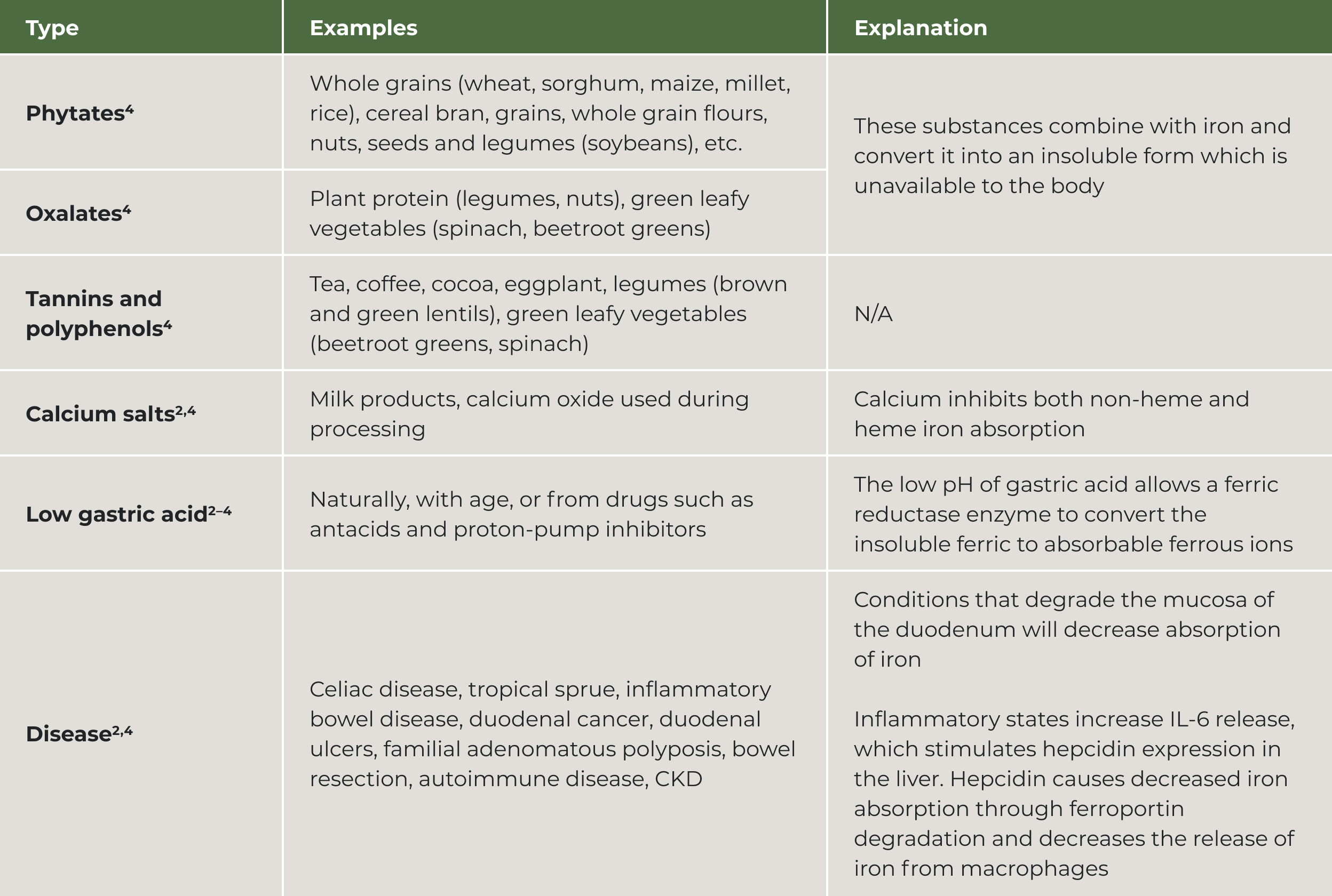
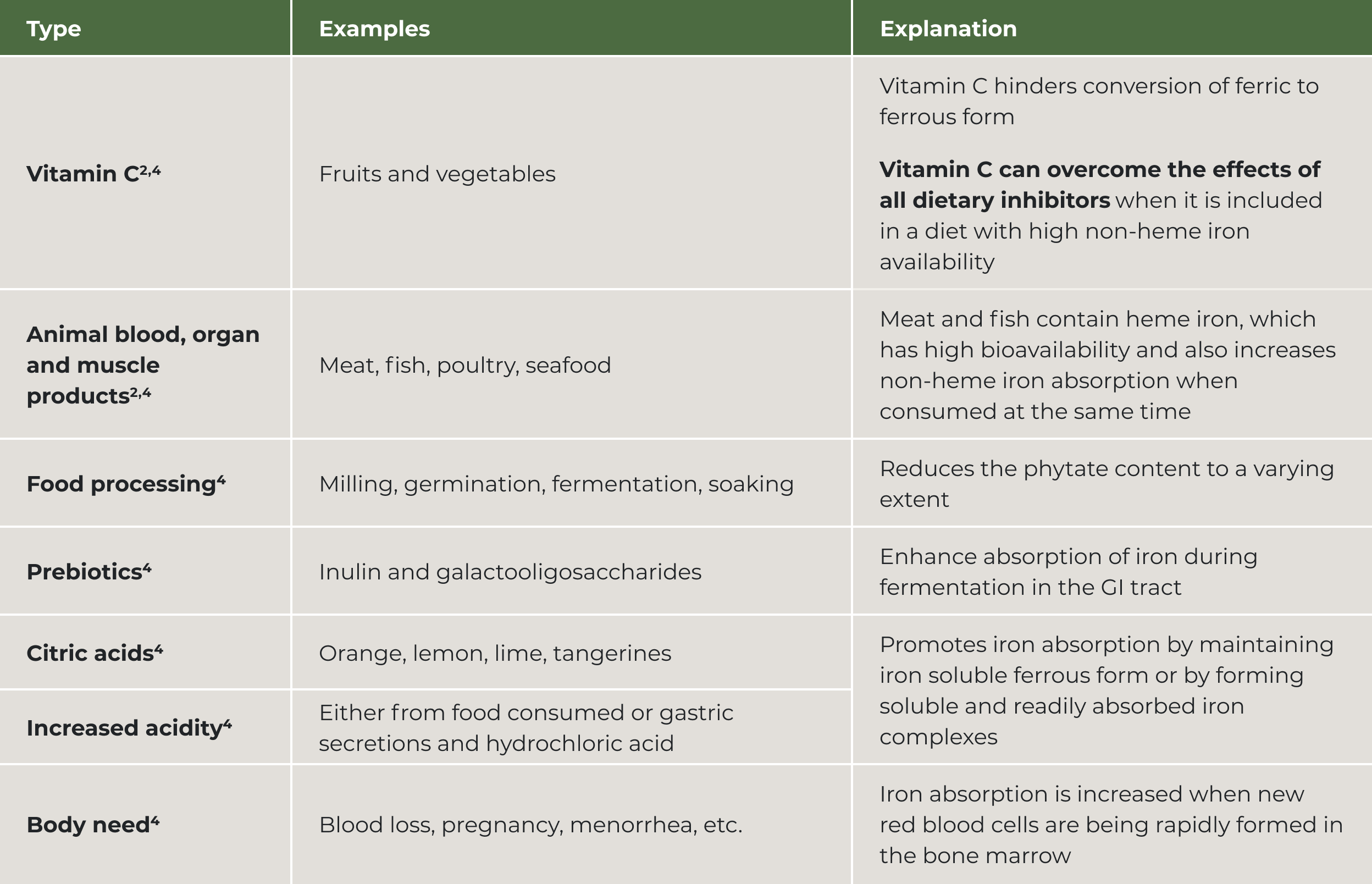
Absorption of iron can be influenced by many dietary, pharmaceutical and disease factors.2,4
Do you have a special interest in nutrition?
Detailed additional information can be found in the below sections.
Dietary inhibitors of absorption

Dietary enhancers of absorption
Available iron in circulation
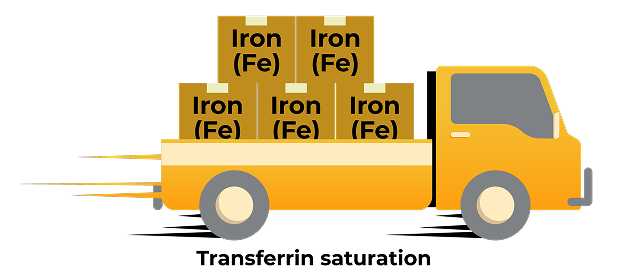
Available iron is transported in the bloodstream to tissues/organs, attached to a protein called transferrin.2
Assessment of available iron in circulation is achieved by measuring total saturated transferrin (TSAT).5
Iron storage
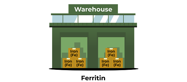
Iron that is not transported upon absorption is stored in enterocytes attached to a protein called ferritin.2
Iron can also be recycled from old or dying red blood cells and stored in splenic or hepatic macrophages.5,6
Iron storage can be assessed by directly measuring ferritin levels.5
Given that disorders in both iron stores and iron distribution can impact iron availability for appropriate RBC production or other functions, “iron deficiency” should be viewed as a mismatch between iron supply and demand, rather than just an indicator of reduced body iron content.7
A state of iron deficiency can therefore occur in the presence of decreased, normal, or increased total body iron.7
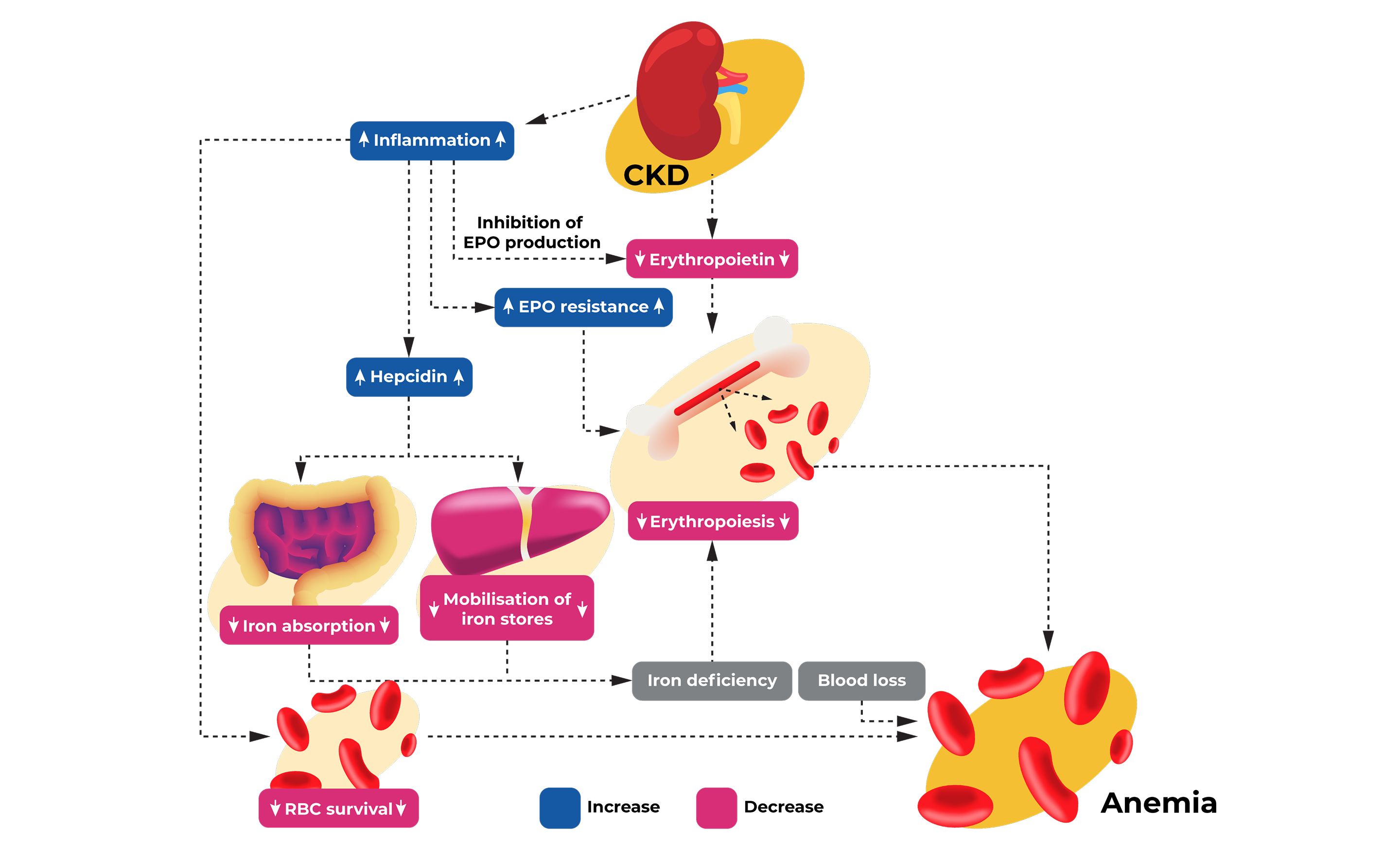
Adapted from Badura K, et al. 2024.8
Hepcidin
Hepcidin is a hormone that acts as a critical regulator of iron homeostasis, or how the body regulates iron levels and metabolism. It is responsible for regulating the absorption of dietary iron through the intestine, in addition to macrophage-managed recycling and re-availability of iron for RBC production or other needs. Increased hepcidin levels in CKD contribute to dysregulated iron homeostasis, or an imbalance in the body’s regulation of iron levels.5
There are many possible tests that can be used to assess iron status. However, KDIGO 2026 guidelines recommend a basic set, including complete blood count (CBC), reticulocyte count, ferritin, and TSAT. Iron deficiency (systemic iron deficiency or inadequate iron availability for erythropoiesis) is then determined using ferritin and TSAT results.5
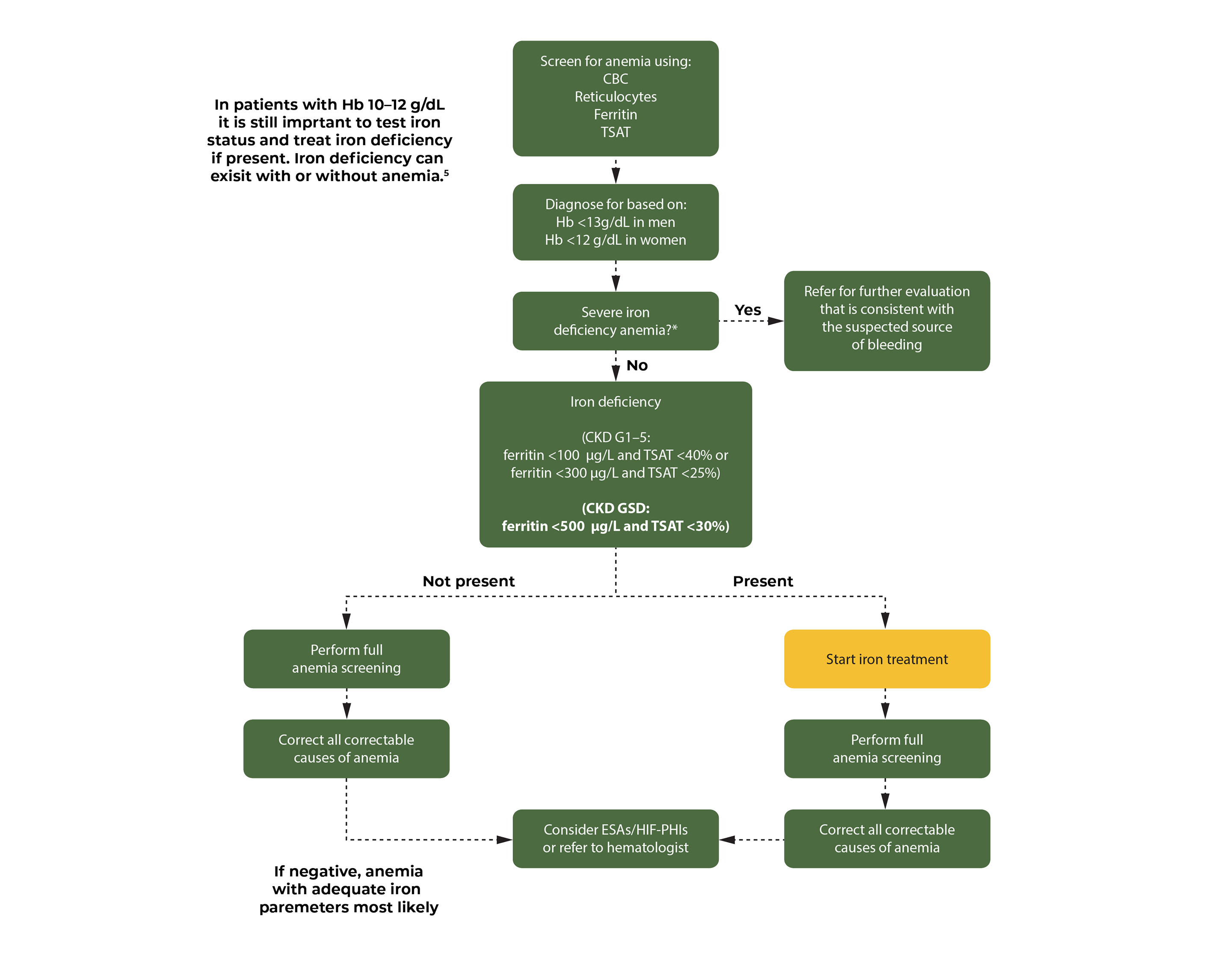
*Severe iron deficiency can be identified by ferritin <45 µg/L or through clinical judgement; OR, if ferritin is unavailable, profound iron deficiency may be identified by a microcytic anemia (MCV <80 fL) in the absence of a known genetic cause.
Adapted from KDIGO 2026.5
KDIGO recommends that a full anemia screening contains:5
- Peripheral blood smear
- Haptoglobin and LDH
- CRP
- Vitamin B12
- Folic acid
- LFTs
- M-protein, free light chains, and urine Bence-Jones
- TSH
- PTH
- Stool analysis
CBC, complete blood count; CRP, C-reactive protein; GI, gastrointestinal; LDH, lactate dehydrogenase; LFTs, liver function tests; MCV, mean corpuscular volume; PTH, parathyroid hormone; TSAT, total saturated transferrin; TSH, thyroid-stimulating hormone.
Despite the diagnosis of anemia and other treatment options using Hb levels to guide treatment decisions, hemoglobin is not a relevant biomarker when considering iron therapy.5
Therefore, regardless of the Hb level, it is still important to test iron status and treat iron deficiency with or without anemia, if iron deficiency is present.5
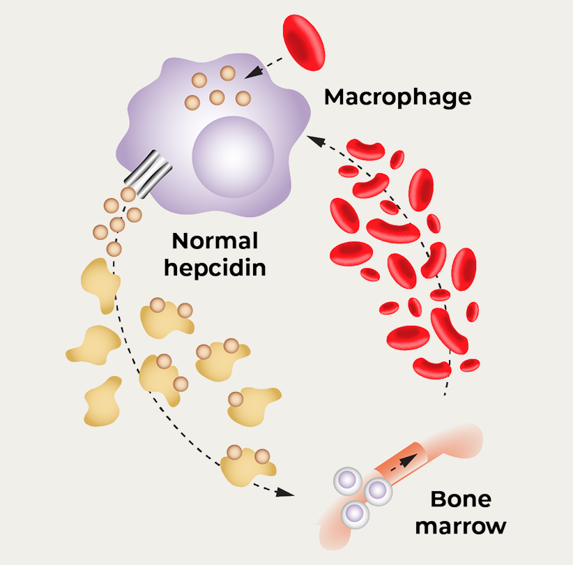
Normal
- Normal iron stores (ferritin)
- Normal iron availability/ supply (TSAT)
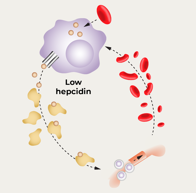
Systemic iron deficiency
- Low iron stores (ferritin)
- Low ferritin (<100 µg/L in non-dialysis CKD patients or ferritin <200 µg/L in CKD G5HD)
- Insufficient iron available for appropriate RBC production
- Low TSAT (<20%)
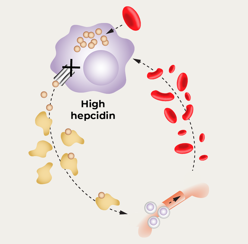
Iron-restricted erythropoiesis
- Normal/High iron stores (ferritin)
- Insufficient iron available for appropriate RBC production
- Low TSAT (<20%)

Adapted from KDIGO 2026.5
Systemic iron deficiency
Historically labeled “absolute iron deficiency”, KDIGO 2026 guidelines propose a change in nomenclature to “systemic iron deficiency” to more accurately reflect the physiological state.5
Systemic iron deficiency is characterized by low TSAT (<20%) and low ferritin level (<100 µg/L in CKD G1–G5 or <200 µg/L in CKD G5HD), reflecting decreased iron levels both in tissue stores and circulation.5
As a consequence of increased losses and decreased absorption, most untreated patients with CKD have reduced iron stores and will progress to a state of systemic iron deficiency unless treated with iron supplementation.7
Iron-restricted erythropoiesis
Historically labeled “functional iron deficiency”, KDIGO 2026 guidelines propose a change in nomenclature to “iron-restricted erythropoiesis” to provide a more physiological representation for why treating people with iron may result in increased erythropoiesis and hemoglobin (Hb) concentration.5
Iron-restricted erythropoiesis is characterized by low TSAT and high ferritin level, reflecting limited iron available for erythropoiesis despite adequate iron stores.5
Even among patients with increased total body iron, reduced iron availability can result in iron-restricted erythropoiesis. For example, in the setting of inflammation, patients can have high ferritin levels, low TSAT, and increased iron stores but still experience restricted erythropoiesis resulting from “reticuloendothelial blockade”.7
Challenges of determining iron status in CKD
Commonly used diagnostic thresholds for ferritin and TSAT, such as ferritin <100–200 μg/L or TSAT<20% do not correlate well with bone marrow iron or Hb response to iron in people with CKD.5
For example, in the general population, a ferritin level <30 μg/l has a high sensitivity and specificity to define iron deficiency. However, in settings where inflammation is common (as in people with CKD), higher cut-offs are chosen as ferritin is an acute phase reactant that increases due to cellular damage and inflammation.5
Nonetheless, because TSAT and ferritin levels are the most commonly used parameters worldwide, are readily available, and are the main parameters utilized in clinical outcome trials to date, they are still recommended to guide diagnosis and management of iron deficiency and anemia associated with CKD.5




References

References
- Warner MJ, Kamran MT. Iron Deficiency Anemia. In: StatPearls [Internet]. Treasure Island (FL): StatPearls Publishing; Updated August 2023. Available from: https://www.ncbi.nlm.nih.gov/books/NBK448065/ Accessed: June 2026.
- Ems T, St Lucia K, Huecker MR. Biochemistry, Iron Absorption. In: StatPearls [Internet]. Treasure Island (FL): StatPearls Publishing; Updated April 2023. Available from: https://www.ncbi.nlm.nih.gov/books/NBK448204/ Accessed: June 2026.
- Camaschella C, Nai A, Silvestri L. Iron metabolism and iron disorders revisited in the hepcidin era. Haematologica. 2020;105(2):260–272.
- Malhotra U, Roy M, Sontakke M, et al. A recent paradigm on iron absorption, prevalence, and emerging dietary approaches to eradicate iron deficiency. Food Bioeng. 2023;2(1):53–63.
- Kidney Disease: Improving Global Outcomes (KDIGO) Anemia Work Group. 2026;109(1S):S1-S99.
- Slusarczyk P, Mleczko-Sanecka K. The Multiple Facets of Iron Recycling. Genes (Basel). 2021;12(9):1364.
- Wish JB, Aronoff GR, Bacon BR, et al. Positive Iron Balance in Chronic Kidney Disease: How Much is Too Much and How to Tell? Am J Nephrol. 2018;47(2):72–83.
- Badura K, Janc J, Wąsik J, et al. Anemia of Chronic Kidney Disease-A Narrative Review of Its Pathophysiology, Diagnosis, and Management. Biomedicines. 2024;12(6):1191.